
Stanford Shared Facilities Select Rayni to Support Their Researchers
Published November 3, 2026
"Rayni's application in the Cell Sciences Imaging Facility at Stanford is to facilitate rigorous and reproducible data generation in the facility. The application is an important part of the integrated user experience that melds expert in person training and support by staff with 24/7 AI intelligence and guidance. The combination provides a constant supporting presence for our users for best practices in instrumentation usage and data generation."
Gordon Wang, Director, Cell Sciences Imaging Facility. Clinical Associate Professor, Psychiatry and Behavioral Sciences.
The Shriram Cell Sciences Imaging Facility, the Neuroscience Microscopy Service, and the Cell Sciences Imaging Facility are shared imaging facilities at Stanford, open to hundreds of researchers across the university. The instruments include:
- Zeiss LSM 780, 880, 900 and 980 confocals
- Zeiss Elyra 7 Lattice SIM
- Zeiss Lattice Lightsheet 7
- Zeiss Axio Observer 7 and Axio Imager widefield
- Zeiss Axioscan Z1 slide scanner
- Leica Stellaris confocal with FLIM and two-photon
- Leica Stellaris 8 DIVE multiphoton
- Bruker TruLive3D light sheet
- Bruker NanoWizard AFM
- Nikon CREST spinning disk
- UltraMicroscope light sheet
Coverage spans the three facilities and keeps growing as each one brings more of its documentation across.
The knowledge and expertise to use these instruments to their fullest extent, and to do so safely while following the documentation and procedures each facility recommends, are the most essential parts of working in a shared facility.
Gordon Wang, Director of the Cell Sciences Imaging Facility, brought in Rayni to make that knowledge accessible right at the instrument. A researcher with reserved time can chat with Rayni's AI to get answers to any question or concern about using the instrument. That helps them complete their task the right way within the limited time of their appointment, avoid misuse or damage, and follow the facility's recommended guidelines.
Answers at the instrument, drawn from each facility's own documentation
Each instrument carries a QR code. A researcher can point a phone camera at it, or open the Rayni AI website on a laptop or computer, and arrive in a conversation about that exact instrument, already loaded with its documentation.
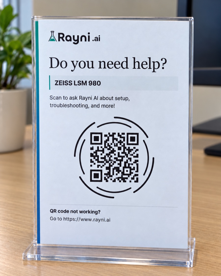
Answers lean on documentation and videos from the lab's leadership, and cite the page they came from, down to the timestamp inside a video. Each facility decides how the platform handles sensitive ground:
- Researchers see warnings and cautions whenever a step is a sensitive one.
- Content marked expert-only stays visible to lab leadership alone, so the riskier material stays with the people trained on it: swapping parts of the instrument, or steps that can lead to downtime.
- The AI points a researcher to the lab manager for sensitive operations, such as anything that touches the instrument's hardware.
- The facility's own documentation takes priority over vendor recommendations.